
In Utero Intra-Amniotic Injection of Poly (lactic-co-glycolic acid) Microparticles Induce Tissue Coverage in the Retinoic Acid Induced Myelomeningocele Rat Model
Nathan L. Maassel1, Douglas Wu2, Nicholas Yung1, Tory Bauer-Pisani4, Mary Elizabeth Guerra1, Sarah J. Ullrich1, Mark Saltzman3, David Stitelman4
1Department of Surgery, Yale School of Medicine, New Haven, Connecticut, United States, 2Case Western Reserve School of Medicine, Cleveland, Ohio, United States, 3Department of Biomedical Engineering, School of Engineering and Applied Science, Yale School of Medicine, New Haven, Connecticut, United States, 4Department of Surgery, Division of Pediatric Surgery, Yale School of Medicine, New Haven, Connecticut, United States
Objective. Determine the effects of intra-amniotic injection of growth-factor loaded microparticles on soft tissue coverage in the rat model of myelomeningocele (MMC).
Design. The rat model of MMC was generated using previously described methods. On gestational day 17, poly (lactic-co-glycolic acid) (PLGA) microparticles (MPs) were delivered to the intra-amniotic space of individual fetuses. Fluorescent PLGA MPs were used to determine rates of particle binding to the MMC defect at 3 hours. Following 3-hour binding experiments PLGA MPs loaded with DiO, bovine serum albumin (BSA), or fibroblast growth factor (FGF) were injected at varying doses, followed by term harvest (gestation day 21, E21).
Setting. Yale animal facility.
Subjects. Sprague Dawley rats.
Interventions. Intra-amniotic injection of MPs
Main outcome. Binding of fluorescent PLGA MPs to MMC defects at 3 hours. Gross and histologic evidence of soft tissue coverage of MMC defects at term.
Results. For 3-hour binding studies, 4 dams were utilized, and 40 fetuses injected. 94% of fetuses with MMC had specific binding of fluorescent MPs to the MMC defect at 3 hours. For term experiments, there were 29 dams, with 187 fetuses injected. Gross soft tissue coverage was apparent in 31% of PLGA-DiO, 55% of PLGA-BSA, and 83% of PLGA-FGF specimens. Histologic coverage ranged from thick particle-based scaffolding, to thin skin-like tissue coverings (Figure).
Conclusion. PLGA microparticles induce soft tissue coverage in the rat model for MMC. Higher incidence of coverage was seen with FGF loaded particles; however, all subtypes had some evidence of coverage suggesting the particles themselves have therapeutic properties for tissue ingrowth.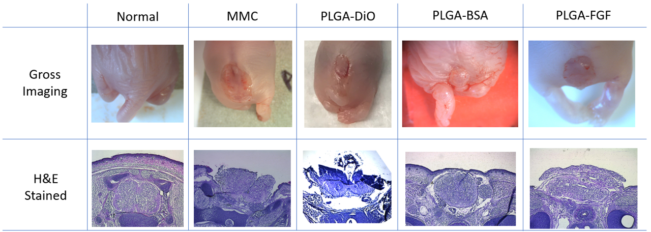 Figure. Gross and histologic imaging of representative term (gestational day 21) rat fetuses. Columns define the experimental groups which are normal (from a non-MMC model dam), MMC (un-injected), PLGA-DiO (fluorescent particles), PLGA-BSA (inert protein loaded particles), PLGA-FGF (growth-factor loaded particles).
Back to 2021 Abstracts